Cardiac Tests
In order to complete an examination of the heart, a number of investigations may have to be performed.
Most of these investigations can be performed as an out-patient, but coronary angiography and the treatment of narrowed coronary arteries will require admission to hospital.
Stress Electrocardiogram
A Stress Electrocardiogram or Exercise ECG is performed while the patient is exercising on a treadmill or stationary bicycle. Symptoms of chest pain or changes on the electrocardiogram after effort can diagnose coronary artery disease. The electrocardiogram is monitored during and after effort and blood pressure readings recorded. The test is stopped if the patient develops chest pain or other symptoms.
Trans-thoracic Echocardiography
Echocardiography of the heart using ultrasound is performed in a similar manner to that of a scan of an unborn baby during pregnancy. The ultrasound produces an image of the heart muscle, and may indicate previous damage to the muscle caused by coronary artery disease or other diseases of the heart muscle. It also provides detailed imaging of the heart valves, providing information about leaking or narrowed valves.
Carotid Ultrasound
Carotid ultrasound can be used to determine whether there are narrowings in the carotid arteries which supply blood to the brain.
The ultrasound probe is placed on the skin overlying the arteries in the neck while the patient lies on his back. Changes in the thickness of the artery sometimes predict similar changes that may occur in the heart. The presence of plaque in the carotid arteries may help decide which patient should be on statin therapy.
CT Coronary Angiogram
This is done as an out-patient procedure by radiologists at the clinic. It is an alternative to invasive coronary angiography and provides a view of the coronary arteries, without the patient having to be admitted to hospital. The test is usually performed when the cardiologist suspects that the heart may be normal, or that there may be only very minor disease present. Prior to this test, blood tests assessing the function of the kidneys should be performed, as the contrast (dye) given during the procedure can aggravate patients with prior renal disease.
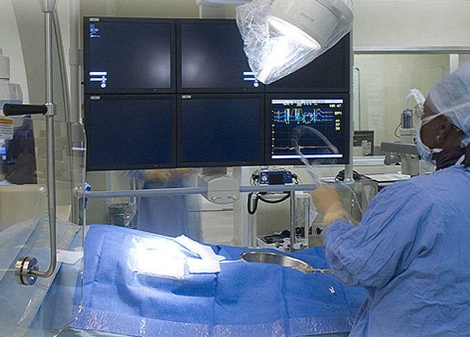
Coronary Angiogram
Coronary Angiography also called cardiac catheterisation, is a detailed way of examining the coronary arteries and is usually performed if the exercise test is positive, or if the patient has typical angina pain. It is the best way to look at the coronary arteries.
The patient will require admission to hospital and is asked not to eat or drink for 6 hours prior to the procedure. Local anaesthetic is injected into the skin above the area of insertion, to numb the area.
It should be noted that this test can be performed from the groin or from the arm.
The catheter is then passed from the relevant area (see animation) in this case from the groin. The catheter is passed up the aorta to the coronary arteries and contrast (iodine) is injected into the arteries. Pictures are taken in various projections which provide very accurate assessment of the state of the arteries.
If narrowings are found, arteries can be stented and opened at the time of the procedure. The procedure is generally very safe. Occasionally patients are allergic to the contrast and this can be adequately dealt with.
Any procedure which involves invasion of the heart does carry risks, but these risks are minimal. The advantages certainly outway the disadvantages.
The main advantage of coronary angiography is the ability to deal with problems at the time of the procedure, that is if narrowings are found, arteries can be stented and opened at the time of the procedure.
Ambulatory ECG Monitoring
Occasionally patients present with palpitations which cannot be recorded in the consulting room. An electrocardiogram recorded over a period of 24, 48 or even 72 hours can be performed by attaching electrodes to the patient. The wires then lead to a small recorder worn on the belt around the patient’s waist.
Showering is a problem during this procedure and has to be avoided while the device is attached. The patient should keep a diary of any symptoms, so that the doctor can correlate any changes that occur on the monitor with this patient’s symptoms.